


home
Home
>
Upper All-On-4 to Replace Failing Bridges and Asymmetric Gums
Upper All-On-4 to Replace Failing Bridges and Asymmetric Gums
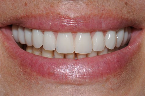
AFTER
This 41-year-old lady had dental treatment done overseas as seen in the before shot. She was not satisfied with the appearance but didn’t really know what to expect. Soon after her return to Australia, she developed pain from numerous of her upper teeth and when seen at our clinic, we discovered the presence of decay affecting many of the teeth under the bridges that were fitted abroad to point that it was not possible to save most of the teeth.
We considered saving 2 teeth, the upper left lateral incisor and canine because they were reasonably healthy, but doing so would have meant that we had to fit an implant-supported bridge segment on either side. This treatment relies on the natural gums for aesthetics, but since the gums here were not only unevenly receded and asymmetrical, but also visible on smiling, the overall aesthetics would have been highly compromised.
Instead, the patient decided to sacrifice two teeth that were otherwise healthy and proceed with All-On-4 dental implants treatment in the top. A feature of this treatment is Aesthetic Gum Replacement, which essentially means that the gum component is replaced as part of the bridge. As such, we had better control and flexibility to design a more balanced look in terms of the appearance of both the teeth as well as the gums, and we were also able to improve the bite at the same time.
Treatment Process
Failing bridges and recession of the gums as a result of gum disease and tooth decay under the upper bridges. Note the collapsed bite seen as a dipping on the right of the picture.
After all, teeth were removed (except one in the very back), the gum line was leveled to allow for aesthetic gum replacement for improved appearance of the gum line, to be incorporated into the design of this All-On-4 Dental Implants prosthesis. Note the straighter alignment and bite.
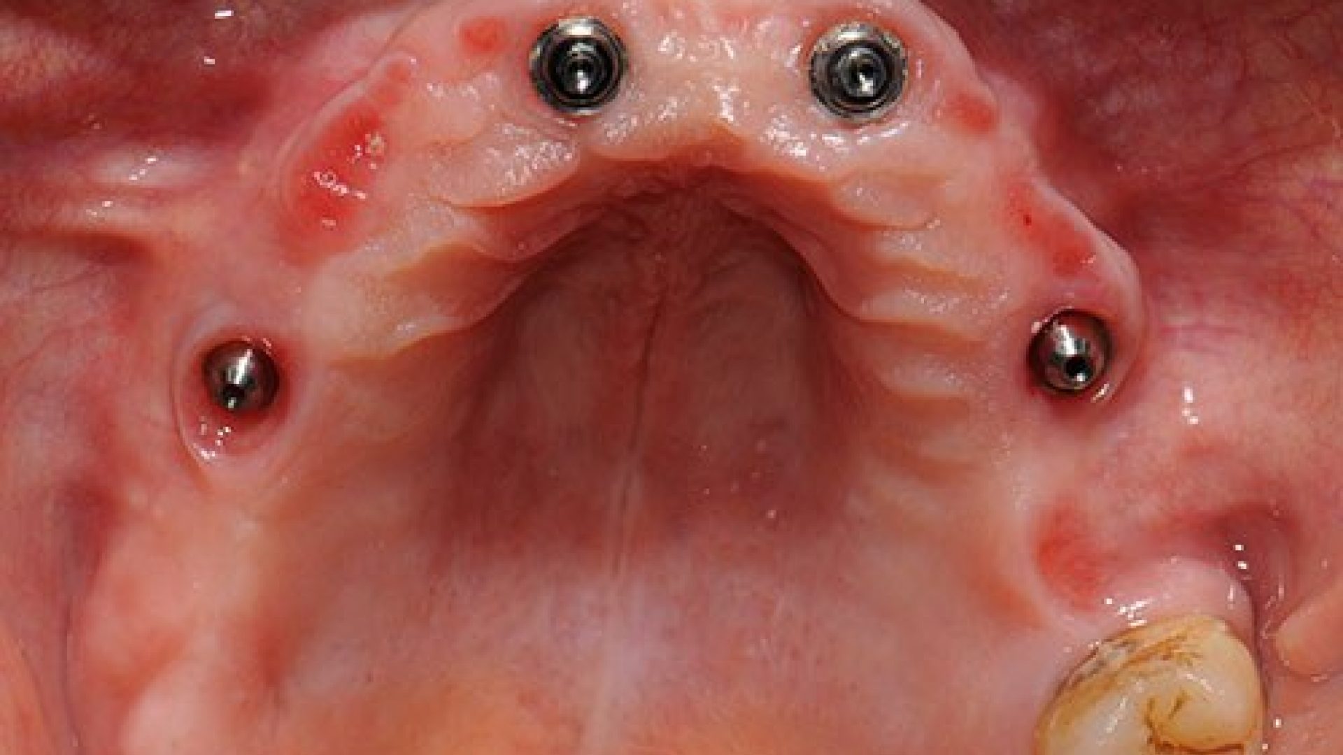
When the upper bridge is removed, the four supporting implants are visible, and the gums under the bridge appear completely healthy despite this patient's history of gum problems with her natural teeth in the past.
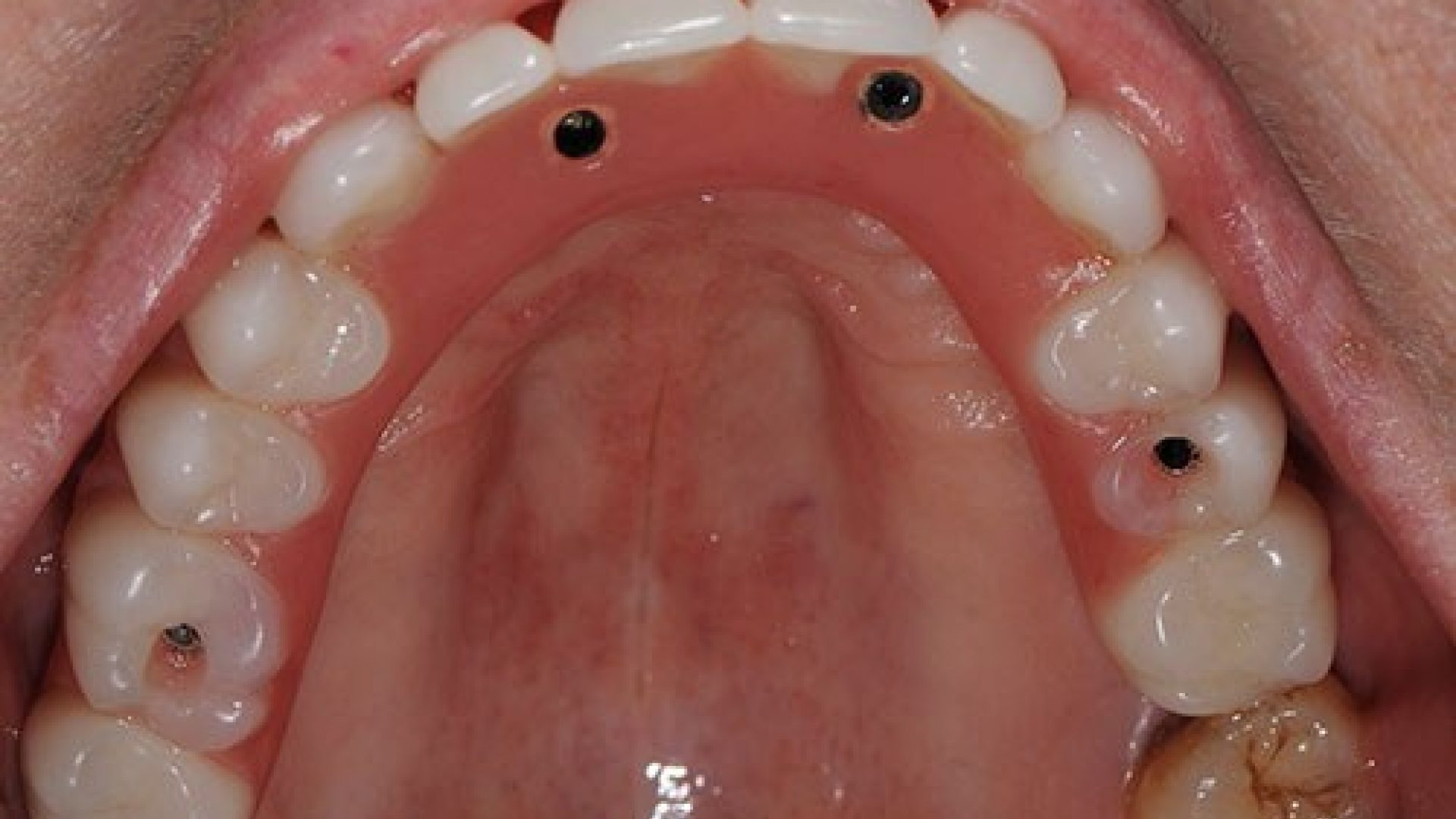
This picture shows the bridge in place over the implants. The holes allow access to the screws to enable removal of the prosthesis when required. These holes get closed over with a coloured material.
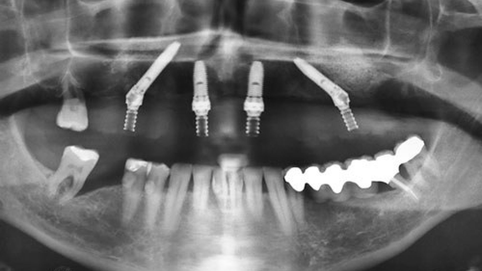
An x-ray that takes 15 months after the surgery illustrates the two angled fixtures in the back and two straight implants in the front, with healthy bone levels.
keyboard_arrow_left
Back to Before & Afters